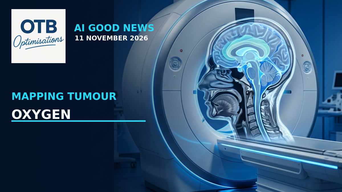
Could a clearer picture of oxygen inside a tumour help doctors decide who needs intensive radiotherapy and who might safely receive less? British researchers are developing an MRI-based approach that could help answer that question.
The Institute of Cancer Research, London, announced the next stage of HYPOXiMAP on 10 November 2026. Working with University College London and the University of Manchester, the team will refine tumour oxygen mapping, including through AI, and test its consistency across hospitals.
This is progress towards better-informed treatment research. It is not yet proof that reducing radiotherapy using these maps preserves cure rates or prevents long-term side effects.
Why Oxygen Matters
Tumours are not biologically uniform. Some regions have less oxygen than others, a condition called hypoxia. Low-oxygen cancer tissue can be more resistant to radiotherapy, making the distribution of oxygen clinically relevant.
HYPOXiMAP uses advanced MRI to make this information easier to see. The research team describes colour maps in which blue marks hypoxic tissue and yellow indicates better-oxygenated regions that typically respond more readily to radiotherapy.
These maps represent information about tissue oxygenation. They are not photographs showing every resistant cancer cell, and a colour alone does not decide an individual patient’s treatment.
Existing Imaging Work and a New Development Programme
According to the ICR, the researchers have already shown that the MRI method can distinguish oxygen-rich from oxygen-poor HPV-positive throat cancers and monitor changes in tumour hypoxia during radiotherapy.
That is an important foundation, but it answers an imaging question rather than establishing the safety of a new treatment strategy. Measuring a biological feature and proving that changing treatment on the basis of that measurement helps patients are different steps.
A £1.63 million Cancer Research UK award will fund a four-year programme to prepare HYPOXiMAP for larger clinical studies. The original ICR announcement confirms this funding, which is not stated in the shorter Medical Xpress version of the story.
Where AI Fits
The planned next phase will use AI to improve the speed, accuracy and consistency of tumour oxygenation measurements. The role is focused: helping refine an imaging process, rather than replacing the people responsible for interpreting scans and planning treatment.
The programme includes a prospective, multicentre study managed by the ICR’s Clinical Trials and Statistics Unit. It will test how consistently HYPOXiMAP performs across different hospitals and MRI scanners.
That matters because a method that works in one specialist setting may behave differently with other equipment, scanning protocols or patient groups. Wider testing is part of establishing a dependable tool, not a formality after an outcome has already been proved.
The Hope Is Gentler Treatment, but the Evidence Must Follow
Radiotherapy can be effective against HPV-positive oropharyngeal, or throat, cancer, but treatment can leave persistent problems such as swallowing difficulties, dry mouth and taste changes. The long-term ambition is to identify patients for whom less intensive treatment could be appropriate without compromising cancer control.
Existing NHS MRI infrastructure makes the approach potentially more accessible than specialised imaging available only in selected research centres. However, the presence of an MRI scanner does not mean HYPOXiMAP is already validated or available as routine care at every hospital.
Why This Is Good News
The value lies in connecting a well-established biological challenge with a practical measurement technique and a plan for testing reliability. Future work can then investigate how the information should influence treatment, rather than assuming that a more detailed scan automatically means better care.
For patients, the important promise is more precise evidence about who might need which treatment. For now, the real achievement is support for the careful research needed to move towards that goal.
Sources and Further Reading
- Institute of Cancer Research: New MRI scan colour codes treatment-resistant cancer cells. Published 10 November 2026. Verified original announcement covering HYPOXiMAP, the £1.63 million award, planned AI refinement and multicentre evaluation.
- Medical Xpress: ICR-provided HYPOXiMAP announcement. Published 10 November 2026. A syndicated account supplied by the Institute of Cancer Research, not an independent clinical trial report.