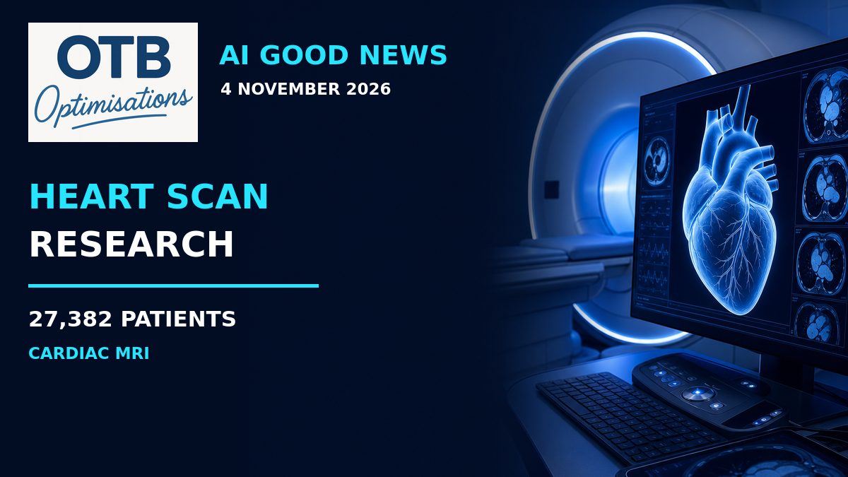
A heart scan contains information that can help doctors understand how well someone’s heart is working. But turning those images into reliable measurements takes time and expertise.
New research co-led by UCL suggests that AI can help with one important part of that process, producing measurements in seconds that were slightly more strongly associated with patients’ subsequent mortality risk than measurements made by experienced clinicians.
The study, announced by UCL on 3 November and published in The Lancet Digital Health, analysed cardiac MRI scans from 27,382 patients in the UK and United States.
It is an encouraging finding about measurement and risk assessment. It is not evidence that AI can predict the exact time of someone’s death, or that using the tool has already improved patient outcomes.
What Is Ejection Fraction?
The researchers focused on ejection fraction, a measurement describing the proportion of blood pumped out of a heart chamber when it contracts.
Doctors use it as one indicator of how effectively the heart is pumping. It can inform decisions about heart-failure treatment, implanted devices and other procedures, alongside a patient’s symptoms, medical history and other tests.
It is also used in assessing surgical risk and monitoring heart function during chemotherapy, according to UCL.
That makes the quality of the measurement important. A useful tool must do more than produce a number quickly: the number needs to be dependable and interpreted in the right clinical context.
A Large Study with Long-Term Follow-Up
The 27,382 patients were undergoing investigation for known or suspected heart conditions. They were followed for an average of more than five years, during which 3,458 deaths occurred.
The team compared ejection-fraction measurements generated by an AI tool with those produced by experienced clinicians assessing the cardiac MRI scans.
The AI-derived measurements were slightly, but statistically significantly, better associated with subsequent mortality risk. UCL reports that the finding remained significant after accounting for factors including age and sex.
This is a retrospective prognostic association: researchers looked at scan measurements alongside later outcomes to see how well the measurements identified differences in risk.
It does not mean the system knew what would happen to every individual. A risk estimate describes uncertainty, not a certainty that a particular person will die or survive.
Seconds for Analysis, Not for the Whole Appointment
The AI tool analysed scans in approximately 2-3 seconds per patient.
UCL compares that with earlier research by the team which found that expert manual analysis typically took around 13 minutes. The manual timing is a comparison drawn from prior research, not a newly reported head-to-head timing measurement in this study.
The distinction matters. These figures concern the analysis task, not the time required to acquire the MRI scan, review the complete case, discuss the result or make a treatment decision.
They do not establish that an entire cardiac appointment now takes seconds. Nor do they demonstrate a reduction in NHS waiting times.
A Small Prognostic Advantage Still Needs Perspective
With a study of this size, a modest difference can be statistically significant. That makes the result worth investigating, but statistical significance does not automatically mean a large benefit for every patient.
The positive result is specific: the AI measurements carried slightly more useful information about mortality risk than the clinician measurements in the analysed data.
The study did not test whether switching patients to an AI-led care pathway saved lives, changed treatment successfully or improved quality of life. Demonstrating those benefits would require additional research.
Why Clinicians Remain Essential
Errors did occur. UCL reports an error rate of around 1% at patient level, affecting 0.08% of images. Those figures describe different units, so they should not be treated as interchangeable.
A low error rate is not a reason to remove human checking. Clinicians still need to verify measurements, recognise when something looks wrong and interpret the scan in the context of the whole patient.
UCL’s senior author Dr Rhodri Davies, who is also the inventor of the tool, emphasises that AI is not replacing clinicians. That relationship to the technology is worth noting alongside the university’s account of the results.
The strongest practical interpretation is a possible first-line measurement tool with expert review where needed, not an autonomous system making decisions about someone’s life.
What Could Come Next?
Faster, consistent measurements could potentially reduce some repetitive analysis work and give clinical teams useful information sooner. Those are possible future benefits, not service improvements demonstrated by this study.
The team plans to explore combining AI heart-scan measurements with information from other tests, particularly electrocardiograms, or ECGs. The aim is to investigate earlier detection of heart-muscle problems.
For now, the good news is a large study supporting a focused use of AI: helping measure heart function quickly, with a small improvement in its association with subsequent risk.
That is a meaningful research step, provided the limits remain clear and people with clinical expertise stay responsible for checking and acting on the results.
Sources and Further Reading
- UCL: AI heart scan measurements better predict survival, published 3 November 2026. Source for the study population, follow-up, deaths, prognostic comparison, analysis timings, error rates and need for human oversight.
- The study in The Lancet Digital Health, linked directly from UCL’s announcement. The summary above follows UCL’s account; access to the journal’s full text may depend on the publisher.
Less hype. More real-world AI progress.